
Definition of Instruction in Teaching and Coaching
Instruction in teaching and coaching both center on structured knowledge transfer, yet differ in delivery․ Teachers design curricula, deliver lessons, and assess learning, while coaches facilitate skill acquisition through guided practice, reflection, and feedback loops, empowering self‑directed growth․ This dual focus ensures learners gain both conceptual understanding and practical competence․ boosting confidence and autonomy․!!
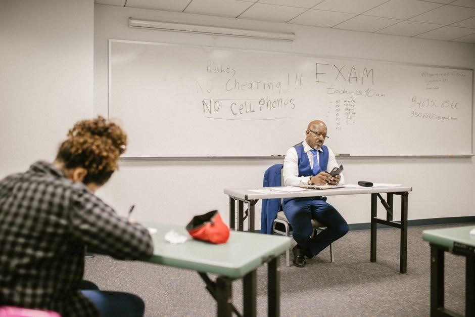
Teaching Involves Structured Curriculum Delivery

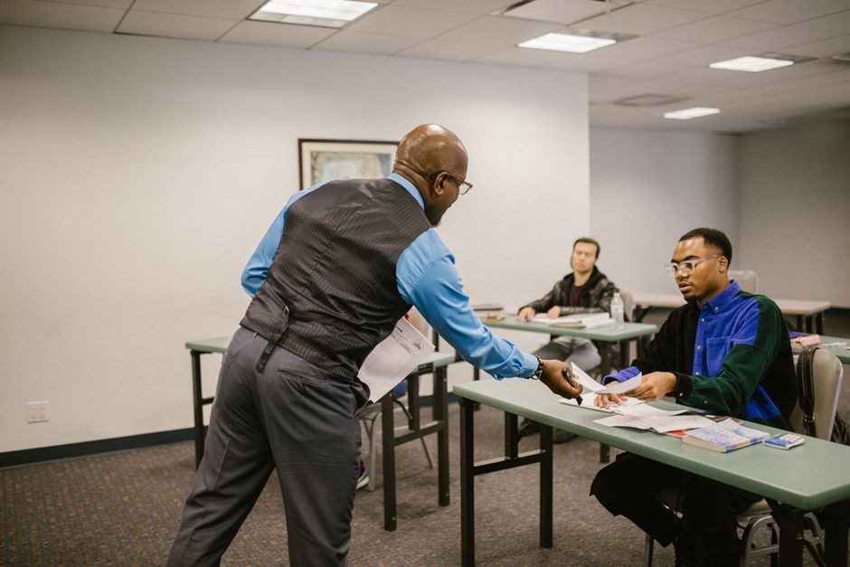
In formal education, instruction is the systematic presentation of content aligned with learning objectives, standards, and assessment criteria․ Teachers design lesson plans that sequence concepts, integrate instructional materials, and employ varied pedagogical strategies—direct instruction, inquiry, collaborative projects—to scaffold from prior knowledge to new concepts․ Timing, pacing, and transitions are mapped to maintain student engagement and ensure coverage of the entire syllabus within the allotted instructional period․
Effective lesson planning begins with clear learning targets, translated into measurable objectives․ Teachers select appropriate resources—textbooks and manipulatives—and design activities that scaffold from prior knowledge to new concepts․ Timing and transitions are mapped to maintain engagement and ensure syllabus coverage․
Assessment practices are interwoven, with formative checks such as quizzes and exit tickets guiding instructional adjustments․ Summative assessments—midterms, projects, and standardized tests—provide accountability and guide curriculum refinement․ By aligning teaching strategies with assessment data, educators foster a learning trajectory that supports masterygrowth․ Steady progress follows!

Coaching Focuses on Skill Development and Self-Directed Learning

Coaching is a collaborative partnership that prioritizes the learner’s autonomy, empowering them to set personal goals, reflect on progress, and take ownership of skill acquisition․ Unlike traditional instruction, coaching refrains from prescribing every step; instead, it asks powerful questions, listens actively, and provides targeted feedback that encourages self‑analysis․ The process begins with a clear, measurable objective, often framed in the learner’s own language, which aligns with broader performance standards but remains personally relevant․
Coaches employ a variety of techniques—modeling, role‑playing, deliberate practice, and reflective journaling—to scaffold skill mastery․ They create a safe space where failure is reframed as a learning opportunity, fostering a growth mindset that sustains motivation․ Through regular check‑ins, coaches track progress using data from practice sessions, self‑assessments, and peer reviews, adjusting strategies in real time․ This dynamic, data‑driven approach ensures that instruction remains responsive to the learner’s evolving needs․
Moreover, coaching emphasizes the transfer of skills across contexts․ By encouraging learners to apply techniques in varied scenarios, coaches help internalize procedural knowledge, turning isolated practice into adaptable expertise․ The result is a learner who not only performs tasks competently but also understands the underlying principles, enabling continuous improvement beyond the coaching relationship․
Ultimately, coaching transforms learning into a lifelong habit, equipping individuals to adapt, innovate, and excel in any evolving world daily today․

Core Similarities Between Teaching and Coaching
Both roles share a commitment to knowledge transfer, motivation, and reflective practice․ Teachers and coaches design learning experiences, assess progress, and adjust strategies to meet learner needs, fostering growth skill development across contexts․
Knowledge Transmission in Both Roles
In both teaching and coaching, the core function is the deliberate transfer of expertise from a more experienced individual to a learner․ This process is structured around clear objectives, contextual relevance, and active engagement․ Teachers design lesson plans that scaffold concepts, employ varied instructional strategies, and use formative assessments to gauge understanding․ Coaches, meanwhile, focus on skill acquisition through modeling, guided practice, and immediate feedback․ Both rely on evidence‑based resources, such as research articles, best‑practice guidelines, and data analytics to inform content choices․ They also create environments that encourage questioning, reflection, and iterative improvement․ The shared emphasis on purposeful communication, alignment with learner goals, and continuous monitoring ensures that knowledge is not merely presented but internalized and applied․ By integrating theory with practice, both educators and coaches facilitate deeper comprehension, foster critical thinking, and promote autonomy, ultimately leading to sustained growth and performance enhancement․ Such synergy between teaching and coaching cultivates holistic learning where curiosity thrives, and growth becomes enduring for allnow․ By intertwining reflective dialogue, evidence‑based strategies, and personalized goal‑setting, educators and coaches jointly nurture resilience, foster autonomy, and create a dynamic learning culture adapts to evolving challenges and celebrates collective achievement․
Motivation as a Key Element
Motivation drives the learner’s engagement, persistence, achievement․ Teachers foster intrinsic motivation by linking content to real‑world relevance, offering autonomy in task selection, and celebrating progress through constructive feedback․ Coaches cultivate a growth mindset by setting clear, attainable goals, providing timely, specific encouragement, and modeling resilience when challenges arise․ Both roles utilize motivational theories—such as self‑determination theory, expectancy‑value theory, and mastery‑orientation to design experiences that satisfy competence, autonomy, and relatedness needs․ By embedding reflective practices, learners internalize purpose, which translates into sustained effort and higher performance․ Moreover, motivation is reinforced through data‑informed progress tracking, allowing educators and coaches to celebrate milestones and recalibrate strategies․ This continuous loop of goal setting, feedback, and self‑assessment ensures that motivation remains a dynamic, central component of instruction, ultimately leading to deeper learning and skill mastery․ In practice, educators and coaches collaborate to align instructional objectives with measurable outcomes, employing diagnostic tools, reflective journals, and peer observation․ This synergy not only enhances content mastery but also nurtures a culture of continuous growth, where feedback loops inform iterative refinement of both teaching strategies and coaching techniques․ Together, they foster lifelong learning!!

Distinct Instructional Approaches
Teaching relies on structured curricula and assessment cycles, while coaching emphasizes collaborative inquiry and reflective dialogue․ Teaching prioritizes content mastery, coaching focuses on personal growth and autonomy, yet both seek measurable progressdaily․

Coaching vs․ Consulting in Instructional Support
In contemporary educational discourse, the distinction between coaching and consulting has become increasingly salient․ While both roles aim to enhance instructional practice, their underlying philosophies and methods diverge markedly․ Consulting is often characterized by a technical, advisory stance: consultants deliver expertise, interpret data, and recommend specific strategies․ Their interventions are typically short‑term, focused on immediate problem resolution, and may lack sustained follow‑up․ Coaching, by contrast, adopts a relational, collaborative framework․ Coaches work closely with teachers over extended periods, facilitating reflective practice, goal setting, and the development of autonomous instructional strategies․ This process is iterative, data‑driven, and centered on building teachers’ capacity to self‑direct improvement․ The literature underscores that coaching fosters long‑term professional growth, whereas consulting tends to provide isolated solutions․ In practice, educators must discern when a consultative approach is warranted—such as interpreting MAP Growth reports or troubleshooting a particular instructional challenge—and when a coaching model is preferable, especially for systemic change and teacher empowerment․ Thus, the choice between coaching and consulting hinges on the desired depth of engagement, the nature of the instructional issue, and the commitment to sustained capacity building․ Moreover, empirical studies demonstrate that schools adopting coaching models report higher teacher satisfaction and student achievement gains compared to those relying solely on consulting interventions․

Assessment and Feedback Mechanisms
Data‑driven assessment guides lesson plans and skill growth․ Feedback loops—formative checks, reflective notes, and performance reviews—enable educators to iterate, align goals, and sustain improvement․ Continuous evaluation drives instructional success․ daily
Data-Driven Decision Making in Teaching
In contemporary classrooms, data‑driven decision making has become a cornerstone of effective instruction․ Teachers routinely analyze standardized test scores, formative assessment results, and growth metrics such as MAP Growth reports to identify learning gaps and inform curriculum adjustments․ By examining trend lines and individual student trajectories, educators can pinpoint specific content areas that require reteaching or enrichment․ This evidence‑based approach allows for targeted interventions, differentiated pacing, and the allocation of instructional resources where they are most needed․ Moreover, data dashboards provide real‑time feedback, enabling teachers to modify lesson plans mid‑unit and to scaffold learning activities that align with students’ current proficiency levels․ When teachers share data insights with students, they promote transparency and foster a growth mindset, encouraging learners to take ownership of their progress․ Collaborative data meetings further strengthen instructional coherence, as teams discuss patterns, share best practices, and collectively decide on instructional strategies that will elevate student outcomes․ Ultimately, data‑driven decision making transforms teaching from a reactive practice into a proactive, continuous improvement cycle that is responsive to the evolving needs of every learner․ Teachers also use data dashboards to benchmark progress, compare cohorts, and tailor interventions that align with district stateUS standards and benchmarks․ Data‑centric equity for learners every daily․
Performance Coaching and Continuous Improvement
Performance coaching in education is a dynamic, iterative process that blends skill development with reflective practice․ Unlike one‑time consultative sessions, coaching emphasizes sustained growth, where educators set measurable goals, observe practice, and receive targeted feedback; Coaches often employ the 5‑step model—clarify, observe, analyze, plan, and reflect—to structure sessions that align with instructional objectives․ This cyclical approach mirrors the continuous improvement loop found in quality management, ensuring that each coaching cycle builds on prior insights․ Data from classroom observations, student work samples, and assessment results feed into the analysis phase, allowing coaches to identify specific instructional strategies that need refinement․ By fostering a culture of self‑assessment, teachers learn to adjust pacing, differentiate content, and experiment with new pedagogical techniques․ The coaching relationship also cultivates professional agency; educators become co‑designers of their instructional practice, leading to increased ownership and sustained motivation․ Over time, accumulation of coaching cycles produces a portfolio of evidence that demonstrates progress, informs professional learning communities, supports district‑wide accountability initiatives․ In essence, performance coaching transforms instruction from a static transmission of knowledge into a living, evolving practice that continuously adapts to student needs and educational standards․

Professional Development and Capacity Building
Instructional coaching blends data insights with reflective practice, guiding teachers to set goals, analyze lessons, and refine strategies․ This fosters continuous improvement, expanding classroom effectiveness and professional confidence ongoing!!․
Instructional Coaching as Capacity Building for Teachers
Coaching also promotes reflective practice, prompting teachers to record observations, set measurable goals, engage in cycles․ Structured coaching sessions follow a cycle of inquiry, data analysis, and action planning, ensuring each intervention is research‑based and tailored to classroom realities․ Over time, this scaffolding not only refines instructional techniques but builds a professional identity rooted in lifelong learning, collaboration, and evidence‑based decision making; Embedding coaching within school culture signals commitment to growth, creating a virtuous cycle where teachers feel supported, valued, and empowered to innovate․ This collaborative framework fosters resilience, ensuring teachers continually refine their craft․!!! Such a framework not only elevates classroom practice but also creates a supportive ecosystem where educators thrive together․ By integrating coaching into everyday practice, schools nurture a culture where continuous improvement and educators feel empowered to experiment, reflect, and grow their students․ for all learners daily!